Correct Art-based Question Chapter 4 Question 8 Part A The structures identified by the arrows are __________. Basic anatomy of the skin Drag the appropriate labels to their respective targets.

A P 1 Lab Ch 7 Hw Flashcards Quizlet
Sweat glands hair follicles dermis sebaceous glands.

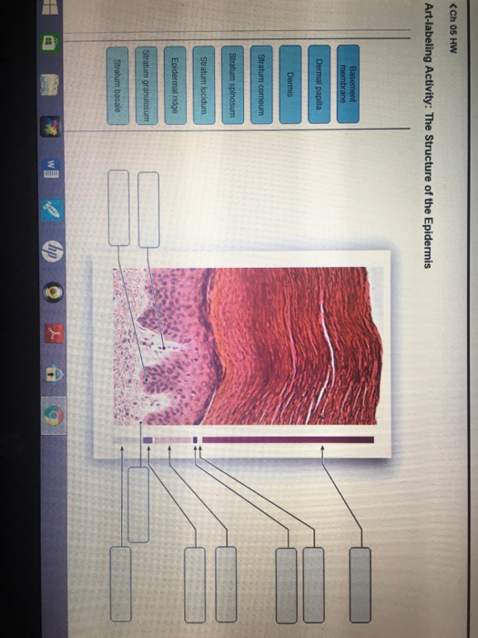
. Correct Chapter Test - Chapter 5 Question 1 Part A Which of these is NOT an accessory structure of the skin. Reticular layer of dermis 5. The Structure of the Epidermis Identify the epidermal layers.
8 rows Art-labeling activity. The structure of the epidermis. Learn vocabulary terms and more with flashcards games and other study tools.
The structure indicated by label E is part of which of the following. Label the integumentary structures listed below on the model of the skin Page 1. Learn vocabulary terms and more with flashcards games and other study tools.
The epidermis consists of five layers of cells each layer with a distinct role to play in the health well-being and functioning of the skin. The Integumentary System Art-labeling Activity. Melanocyte in the Stratum Basale of the Epidermis.
Epithelial root sheath Epithelial root sheath Dermal root sheath Matrix Arrector pili muscle Hair follicle Hair bulb Hair papilla bFrontal section of a hair root and hair follicle Cortex. The epidermis regenerates in orderly fashion by cell division of keratinocytes in the basal layer with maturing daughter cells becoming increasingly keratinised as they move to the skin surface. Lab - Integumentary System 226 Correct Art-Labeling Activity.
Start studying Art-labeling Activity. Part A Drag the labels onto the epidermal layers. The thickness of skin varies from 05mm thick on the eyelids to 40mm thick on the heels of your feet.
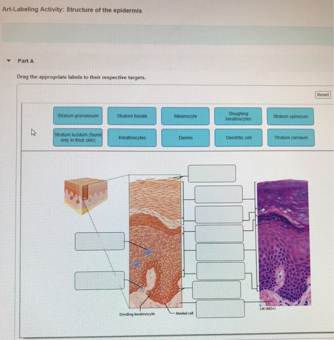
Part A Diagram of. The Structure of the Epidermis Stratum cormeum Stratum basale Dermal papilla Epidermal ridge Stratum lucidum Stratum granulosum Basement membrane Stratum spinosum Dermis. Observe where the basement membrane separates the epidermis and dermis.
Correct Intercalated disks are found between cardiac muscle cells and allow them to communicate and contract in unison. Structures of the epidermis epidermal ridge stratum lucidum stratum corneum tactile discs. The protein found in large amounts in the outermost layer of epidermal cells is collagen.
Use the histology images to label nail structures Page 2 and hair structures Page 3. The epidermis is a dynamic structure acting as a semi-permeable barrier with a layer of flat anuclear cells at the surface stratum corneum. Biology Chemistry Earth Science Physics Space Science View all.
Correct Stratified squamous epithelium like that found in the. What structure is responsible for increasing surface area to provide for the strength of attachment between the epidermis and dermis. Structures of the Epidermis-Dermis Junction 2 of 79.
Skin that has four layers of cells is referred to as thin skin From deep to superficial these layers are the stratum basale stratum spinosum stratum granulosum and stratum. Anatomy and Physiology questions and answers. Thick and thin skin A hypothetical drug causes blood vessels to grow from the dermis into the superficial stratum granulosum of the epidermis.
English French German Latin Spanish View all. Label the skin structures in Figure 71. Which of these is NOT a function of the layer at D.
Layer B is composed primarily of __________. Hair Structure Part A Drag the appropriate labels to their respective targets Reset Help Medulla Melanocyte Hair shaft Dermal root sheath Hair root Cuticle Hair follicle. Structure of the epidermis Part A Drag the appropriate labels to their respective targets.
You identify this tissue as __________. Hypodermis Label the layers of the epidermis in thick skin in Figure 72. Papillary layer of Dermis 4.
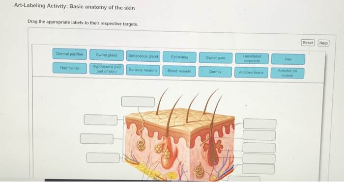
Structure of the epidermis PartA Drag the appropriate labels to their respective targets. Correct Chapter 4 Chapter Test Question 5 Part A You are observing a tissue under the microscope and you see multiple layers of flattened cells. Help Reset Sweat pore Dermal papillae Epidermis Hair Dermis Sebaceous gland Hypodermis not part of skin Hair follicle Blood vessels Sweat gland Sensory neurons Adipose tissue Lamellated.
To supply cells to replace those lost. Start studying Basic Structures of the Epidermis-Dermis Junction. What effect would you see in the most superficial epidermal layers.
Draw a bracket the identifies each stratum of the epidermis Page 4. Help Reset Lamelated Dermal papillae Sweat gland Sebaceous gland Hair Epidermis Sweat pore corpuscle Hypodermis not part of skin Arrector pil muscle Hair follicle Blood vessels Sensory neurons Dermis Adipose tissue. Structure of muscle tissues Part A nose external ear.
Summary of epithelial tissues Part A Drag the appropriate labels to their respective targets.
Solved Art Labeling Activity Structure Of The Epidermis Chegg Com
Solved Art Labeling Activity Basic Anatomy Of The Skin Drag Chegg Com
Solved Ch 05 Hw Art Labeling Activity The Structure Of The Chegg Com
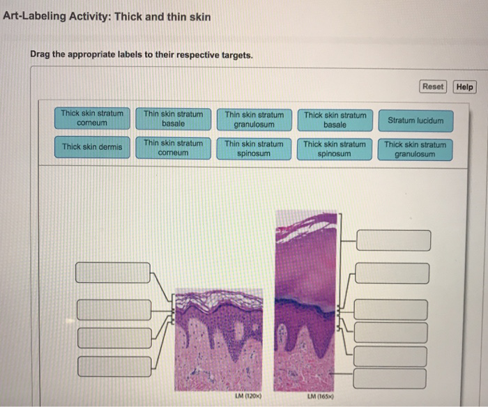
Solved Art Labeling Activity Thick And Thin Skin Drag The Chegg Com

Lab Practice 4 Integumentary System Flashcards Quizlet
Solved Reset Help Hair Hypodermis Not Part Of Skin Adipose Chegg Com

Solved Art Labeling Activity The Structure Of The Epidermis Chegg Com

Chapter 6 Study Set Flashcards Quizlet
0 comments
Post a Comment